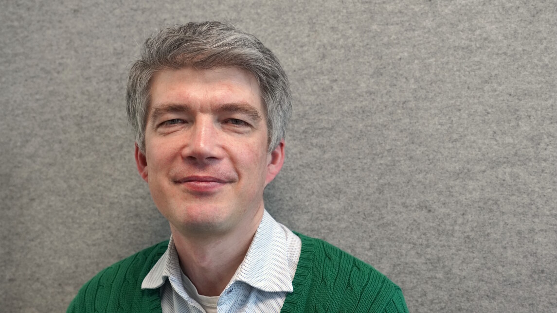
Postdoctoral Researcher David Molnar is conducting a cohort study to identify features of the epicardial adipose tissue that indicate risk of myocardial infarction. He is developing an AI model that can analyse the risk factors apparent in CT scans, enabling more accurate and confident risk assessment by clinicians.
David Molnar, Postdoctoral Researcher at the University’s MSCA-co-funded SYS-LIFE programme, has moved into full-time research from his position as a PET radiologist at Sahlgrenska University Hospital in Gothenburg, Sweden. Molnar thinks his clinical background is beneficial to his research:
“Clinical experience is a great benefit to the research community: in order to produce relevant research, which is sensibly placed in the medical field with regards to clinical applicability and also pathophysiological processes, the clinical eye is important.”
Clinical applicability is at the core of Molnar’s research, as he wants to help clinicians more accurately assess their patients’ risks of myocardial infarction. His research could save lives as well as medication costs by creating a method of classifying high-risk and low-risk individuals.
Molnar’s current research is based upon his PhD, during which he developed an AI model to analyse large image datasets. The model was initially developed for and trained on the Swedish SCAPIS cohort, which includes CT scans of the hearts of 30,000 healthy individuals between the ages of 50 and 64. Now Molnar is developing his AI model further, so that it can be used for detailed analysis of the epicardial adipose tissue of clinical patients.
The epicardial adipose tissue and what it can tell us
The epicardial adipose tissue is a layer of fat surrounding the heart. The thickness and radiodensity of the tissue differ between people based on their bodyweight, as lean people typically have a thinner epicardial adipose tissue, and so forth.
There is no consensus about the exact functions of the epicardial adipose tissue. Molnar names some of the possible functions:
“It serves as a frame for the coronary arteries, which are embedded in and tunnelled through this tissue. It seems to hold them in place and protect them against deformation during heart movements. It also probably serves as an energy reservoir for the heart muscle, as there is no membrane separating it from the heart.”
When the epicardial adipose tissue gets thicker, it also seems to become inflamed, which could be one of the reasons why the fatty depot is so important in the development of coronary artery disease. Molnar wants to have a closer look at the tissue and the coronary arteries to identify features that indicate risk of myocardial infarction.
“We believe that there is information in parts of the fatty tissue which we can discover if we take a more zoomed in picture. So far, we have only looked at the total volume of fat, and we are likely missing some information. We believe that we can further improve the identification of high-risk individuals by looking more locally around the arteries.”
Improving risk assessment with AI
To analyse the features of the epicardial adipose tissue, Molnar is using the AI model he developed during his PhD. The model is used on a cohort from the Turku University Hospital, with an imaging dataset consisting of 3,000 clinical patients with suspected coronary artery disease. Molnar finds the cohort’s images to be a good addition to the earlier SCAPIS data, which has images of healthy, or at least asymptomatic individuals.
Molnar hopes that his AI model can one day be integrated into clinical practice as a support for clinicians assessing patients’ risks of myocardial infarction. The model performs much better than expected on the Turku cohort, even though the image characteristics are different than the SCAPIS images. Molnar is currently developing the model further.
According to Molnar, the practical application of the AI analysis into clinical practice would be easy and cost-effective. Everything the model needs is already available from the standard imaging protocol, which is readily implemented in clinical use globally:
“The clinicians would only need to run this model and analyse the existing images. We could obtain valuable information and help identify the high-risk individuals at no extra cost – neither financial nor in terms of extra radiation.”
By identifying signs of disease, Molnar wants to help clinicians be more comfortable with treating high-risk individuals more aggressively than today. Learning about high-risk features also makes it possible to identify reliable criteria for low-risk patients:
“Among those labelled as high-risk today, we do in fact have a proportion of people who are falsely labelled as high-risk. They will not get a myocardial infarction, and they are treated unnecessarily. We can improve diagnostic precision and help the health care system in the very difficult task of sorting people into different groups – high risk, low risk, medium risk – and to verify diagnosis in a sensible way that benefits the patient.”
SYS-LIFE, Systemic Approaches to Improve Cardiometabolic and Brain Health during Lifespan is Marie Skłodowska-Curie postdoctoral programme cofunded by University of Turku and European union (project 101126611) in 2023–2028. SYS-LIFE supports excellent international early and mid-career stage researchers by providing 22 three-year bottom-up project grants in cardiometabolic and brain research, complemented with training and possibility for secondments outside academia. SYS-LIFE partners include Turku University Hospital, Business Turku, Siemens Healthineers and Ghent University.
Text and photo: Iida Taskila