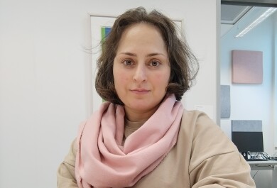
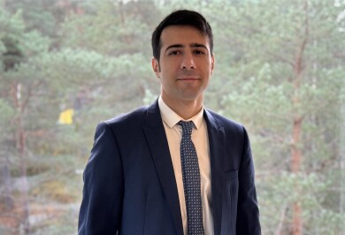
Dissertation defence (Clinical Physiology and Nuclear Medicine): BSc, PharmD, MSc Imran Iqbal
Time
BSc, PharmD, MSc Imran Iqbal defends the dissertation in Clinical Physiology and Nuclear Medicine titled “New Approaches To Molecular Imaging Of Inflammation And Fibrosis In Myocardial Injury” at the University of Turku on 20 November 2026 at 12.00 (University of Turku, Turku School of Economics, Osuuskauppa Auditorium, Rehtorinpellonkatu 3, Turku).
Opponent: Professor Riikka Kivelä, PhD (University of Jyväskylä)
Custos: Professor Antti Saraste, MD, PhD (University of Turku)
Summary of the Doctoral Dissertation:
Heart damage can trigger two harmful processes: inflammation and the build-up of scar tissue. Both can weaken the heart over time and contribute to heart failure. My dissertation investigated whether these changes can be detected non-invasively using positron emission tomography, or PET, before they cause more serious damage.
The research focused on two newly developed imaging tracers. One tracer was designed to detect activated immune cells that gather in damaged heart tissue after a heart attack. The second tracer was designed to detect activated cells involved in the formation of scar tissue in the heart.
In animal models, both tracers were able to identify areas of heart damage. The inflammation-targeting tracer accumulated more strongly in areas affected by myocardial infarction than in healthy regions of the heart. Its uptake was also associated with worsening heart function, suggesting that it may provide useful information about the severity of ongoing inflammation.
The fibrosis-targeting tracer showed higher uptake in areas where scar tissue had developed. This indicates that PET imaging could potentially be used to visualize fibrosis in the heart without relying only on tissue samples or indirect measurements.
The study also examined whether PET imaging could be used to monitor the effects of a potential treatment. A nanoparticle-based therapy was tested to see whether it could reduce the activity of inflammatory cells after myocardial infarction. In this study, the treatment did not produce a measurable reduction in tracer uptake, suggesting that it did not significantly reduce the inflammatory activity detected by PET.
Overall, the main finding of my dissertation is that PET imaging can provide information about two important biological processes involved in heart disease: inflammation and fibrosis. Instead of showing only the structure or pumping function of the heart, these imaging methods can help reveal what is happening at the cellular level inside damaged heart tissue.
The broader importance of this work lies in the possibility of detecting harmful changes in the heart earlier and more precisely. In the future, such imaging methods could help researchers and clinicians identify patients with active inflammation or fibrosis, follow how these processes change over time, and evaluate whether new treatments are working.
Although these studies were performed in experimental animal models and further research is needed before the methods can be routinely used in patients, the findings provide an important step towards more personalized imaging of cardiovascular disease. Ultimately, this approach could support earlier diagnosis, better treatment monitoring, and more targeted care for patients with heart disease.