New Raman imaging microscope reveals structural and compositional variations in materials
Materials Chemistry Research Group (MCRG) at the University of Turku obtained a Raman microscope device. This unique instrument offers information on structure and chemical properties of materials, non-destructively.
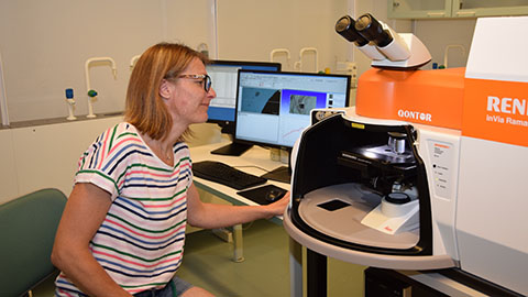
Raman spectroscopy is a powerful analytical tool that can be applied to the analysis of a wide range of materials – from polymers and coating compositions to paintwork and semi-conductive materials. These analyses can be carried out in the MCRG using the new Renishaw inVia Qontor confocal Raman microscope system.
The Raman microscope is equipped with four laser lines, ranging from near-IR (785 nm) to visible (633 and 532 nm) and near-UV (355 nm). It also has a Raman mapping and focus track capability. This microscope is unique in Finland and it was purchased with financial aid from the University of Turku for strategically important research infrastructure (2017) and from funding granted by the University principal for research equipment (2018).
– The new Raman microscope system will play a leading role in the research and development of materials for a wide range of engineering applications in our research group, says Pia Damlin, one of the researchers of the MCRG. As examples for engineering applications Damlin mentions energy storage, photovoltaic applications and different structures produced from carbon.
Theory behind Raman spectroscopy
Vibrational spectra obtained using IR or Raman spectroscopy results from the interaction of the vibrational motions of a molecule with electromagnetic radiation. By studying the vibration of the atoms we can discover the chemical composition of the unknown material as the obtained spectrum is always unique – like a fingerprint – to the material under study.
The above mentioned techniques differ in their way of signal generation. While IR spectra, in a similar way to UV-Vis, are obtained in the absorption mode, Raman spectroscopy is a scattering technique. When a Raman spectrometer is combined with a confocal microscope, it is even possible to visualize the distribution of materials on the micrometer scale and to do depth profiling analysis of structured materials.
Why use Raman?
Due to its sensitivity, high information content, and non-destructive nature, Raman is now used in many applications across the fields of chemistry, biology, geology, pharmacology, forensics, pharmaceuticals, materials science, and failure analysis. Spectral libraries in excess of 16,000 compounds are now available for direct compound identification.
Infrared and Raman spectroscopy are used as complementary techniques in the MCRG because each method looks at different aspects of a given sample. While IR is sensitive to functional groups and to highly polar bonds, Raman is more sensitive to backbone structures and symmetric bonds.
– The newly purchased Raman device makes a perfect and important match to our existing IR equipment. Using both techniques provides twice the information about materials vibrational structure than can be obtained by using either alone, explains Damlin.
How to utilise Raman?
In collaboration with the Department of Archaeology (UTU) the Raman microscope has been used in order to identify mineral pigments from Turku Cathedral wood fragments. Raman spectroscopy is perfect for the analysis of historical relics due to its non-destructive nature. It offers information about origin, authenticity and age, while preserving the whole sample.
Recent advancements in Raman imaging have directly impacted the scale to which carbon nanomaterials can be characterized, enabling the characterization over large sample areas in a short time period. Together with the KTH Royal Institute of Technology in Stockholm cellulose derived carbon nanodots have been analyzed using Raman microscopy. Raman imaging is a valuable tool for applications that require detailed spatial resolution and large-scale quantification of chemical and structural features.
– In our research group Raman spectroscopy will play an important role to facilitate the advancement of cutting-edge nanotechnology research in the future. With the new Raman microscope we are able to take a step further.
Text: Sampo Hirvioja
Photo: Kari Loikas