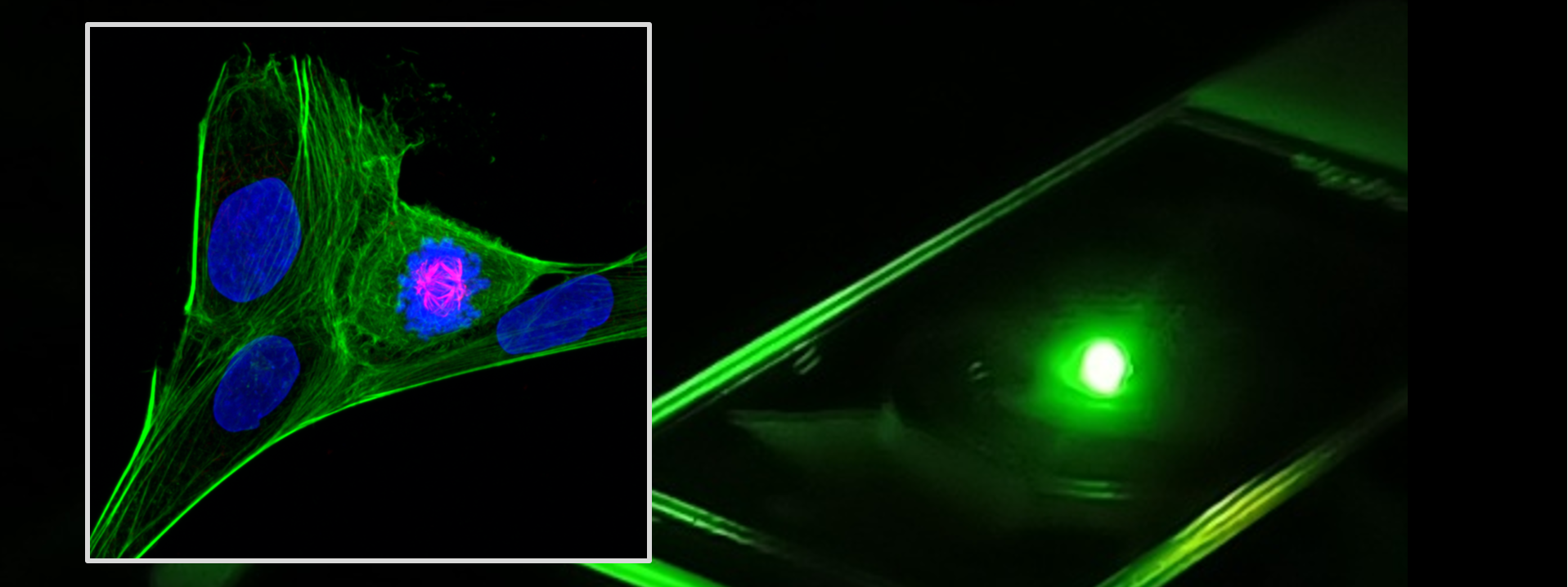
Medisiina Imaging Centre
Medisiina Imaging Centre in the Medisiina C2 provides selection of modern bioimaging devices.
Contact mic@utu.fi to book your training and for more information.
- Instruments are available for all users in academic groups as well as companies.
- Access is given after mandatory user training.
- Instruments are booked in OpenIRIS.
- Some instruments have user fees. The fee is shown in OpenIRIS when booking.
- Contact mic@utu.fi for more information about our instruments and methods.
Main features:
- Objectives: 5x/0.16NA, 10x/0.45NA, 20x/0.8NA, 20x/0.4NA long working distance, 40x/0.6NA long working distance, 40x/1.2 water immersion, 63x/1.4 oil immersion, 100x/1.42NA oil immersion
- Lasers: 405 nm, 488 nm, 561 nm, 640 nm
- Emission filters: 445/45 nm, 525/30 nm, 600/37 nm, 617/73 nm, 692/40 nm, 440/521/607/700 nm quad band
- Prime BSI sCMOS camera
- Holders: slides, 35 mm dishes, wellplates
- Live imaging chamber (temperature and CO2 control)
> Full specifications and the user guide
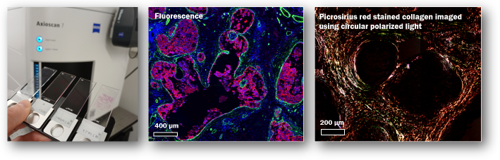
Zeiss Axioscan 7 is a versatile whole slide scanner capable of automatic whole slide imaging in brightfield, fluorescence, and polarized light microscopy. It is equipped for imaging of biological tissue and geological thin sections.
- Objectives:
N-Achroplan 5x/0.15, PApo 20x/0.8, EC Plan-Neofluar 20x/0.5 Pol, EC Epiplan-Neofluar 20x/0.5 Pol, Plan-Apochromat 40x/0.95 - Fluorescence filters:
- Kromnigon SpectraSplit® 7 for 7 channel imaging:
385 nm, 430 nm, 470 nm, 555 nm, 585 nm, 635 nm, and 735 nm - Zeiss single band filter cubes: Filter Set 96 BFP (DAPI), Filter Set 38 GFP, Filter Set 43 Cy3, Filter Set 50 Cy5
- Filter set 90 HE LED (multiband for DAPI, 488, 568, 647)
- Kromnigon SpectraSplit® 7 for 7 channel imaging:
- Fluorescence LED excitation light source:
Zeiss Viluma 7 (model FR-R[G/Y]BV-UV) - Cameras:
- Zeiss Axiocam 705 color
- Hamamatsu Orca Flash4.0 V3 (Monochrome)
- Polarized light microscopy setup:
- Circular polarization
- Rotatable linear crossed polarizers for 0-90 degrees with 15 degree steps
- Slide formats:
- Normal slides 76mm x 26mm , macro slides 76mm x 52mm, geological slides 28mm x 48mm
- Maximum slide thickness is 1.30 mm for biological and 1.60 mm for geological samples.
Operator assisted imaging available for occasional users.
3DHistech Pannoramic slide scanners are used to automatically image whole tissue section slides stained with brightfield or fluorescence stains.
Instruments:
Pannoramic P1000 slide scanner
- Brightfield
- Slide formats: 25 mm x 75 mm and macro slide (50 mm x 75 mm)
- 20x/0.8 and 40x/0.95 objectives
- Capacity: 600 normal slides and 200 macro slides
Pannoramic Midi fluorescence slide scanner
- Fluorescence and brightfield
- 20x/0.8 and 40/0.95 objectives
- Fluorescence filters:
- Capacity: normal slides (25 mm x 75 mm)
Pannoramic P250 slide scanner
- Brightfield
- Objective 20x/0.8
- Capacity: 250 normal slides (25 mm x 75 xx)
Slide scanner output file format is Mirax (mrxs). Install the free CaseViewer for viewing images.
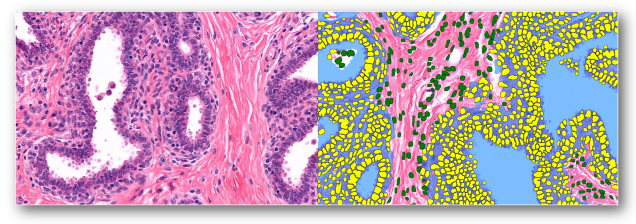
We have two workstations for analyzing whole slide images using Visiopharm software.
- Brightfield and fluorescence images
- Use existing analysis pipelines or develop your own
- Trainable deep learning classifier
- Intuitive graphical user interface. No programming needed.
Bruker Skyscan 1272
- High resolution ex vivo volumetric analysis and three-dimensional imaging of relatively small bone samples
- Imaging of soft tissues is possible if their radioabsorbance differs from surrounding tissues
- Imaging of vasculature can be achieved by perfusion of samples with radiocontrasting agents
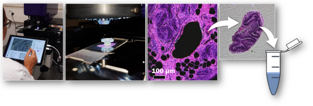
Laser capture microdissection (LCM) is used to cut the areas of interest from tissue sections for different downstream molecular analyses. Our instrument is Acculift by Targeted Bioscience.
Main features:
- Ultraviolet laser (UV) cutting and infrared laser capture (IR)
- Sample types: FFPE, frozen sections, cell cultures
- Area identification by brightfield or fluorescence staining
- Best results are obtained by using membrane slides for preparing the sections. Slides are available from us.
- Dissected tissue is collected using GeckoGrip caps. Caps are also available from us.
- Protocols have been set up by our users to analyze the following from the collected tissue:
- RNA
- DNA
- Proteomics using mass spectrometry
Zeiss AxioImager.M1
- Epifluorescence, brightfield and phase contrast
- Objectives: 5x/0.16NA, 10x/0.3NA, 20x/0.5NA, 40x/0.65NA, 40x/1.3 oil immersion, 63x/1.4 oil immersion
- Filter cubes:
- DAPI (Ex: 359/48 nm, Em: 445/50 nm)
- GFP (Ex: 470/40 nm, Em: 525/50 nm)
- Rhodamine (Ex: 546/12 nm, Em: 607/65 nm)
- Alexa Fluor 660 (Ex:600/50 nm, Em: 685/50 nm)
- Cameras: Zeiss AxioCam MRm (monochrome) and Zeiss AxioCam 506 (color)
- Fluorescence excitation light source: CoolLED pE-300 LED
- Stereo microscope
- Trans- and reflected light illumination
- Camera: Zeiss Axiocam ERc5S
- Polarization, brightfield
- Objectives: 2x/0.10NA, 4x/0.13NA, 10x/0.30NA, 20x/0.50NA, 40x/0.75NA, 60x/0.85NA
- Camera: Nikon DS-Fi3 (color)
- Illumination: CoolLED pE-100white (brightfield)
- Brightfield
- Objectives: 2x/0.05NA, 4x/0.1NA, 10x/0.25NA, 20x/0.4NA, 40x/0.6NA, 60x/1.45 oil immersion
- Camera: Zeiss Axiocam ERc5S
Azure Sapphire Biomolecular Imager
The Sapphire Biomolecular RGBNIR Imager is a next-generation laser scanning system that provides exceptional data quality through extremely sensitive detection, ultra high resolution, and broad linear dynamic range. The Sapphire is capable of fluorescence, chemiluminescence, and phosphorimaging. Fluorescence excitation (nm): 488, 520, 658 and 784.
Fuji BAS-5000
Fuji Analyzer BAS-5000 is a phosphorimager that scans autoradiography plates. It can be used for scanning the image stored on special autoradiography plates that store energy (for example from radioactivity (beta, gamma)) into small crystals on the plate from ex vivo, in vitro, etc. studies. This scanner can reach up to 25 micrometer resolution. The BAS-5000 is no longer manufactured and newer phosphorimagers do not achieve the same scan speed, sensitivity and other characteristics anymore.
Fuji LAS-4000 (membrane and gel imaging)