Imaging Is Advancing in Leaps and Bounds
– We are currently living the second renaissance period of imaging. The first period took place when glass was invented. Now we are creating new, more accurate and smaller technology that tells us more, and are introducing microscopy to disciplines where it has not been used before, says Teng-Leong Chew. Next to him, Pasi Kankaanpää nods approvingly. – On the field, people are talking about a revolution, says Kankaanpää.

According to Teng-Leong Chew and Pasi Kankaanpää, researchers of imaging are engaged in international and multidisciplinary collaboration. The goal is to introduce microscopy to as many areas of research as possible.
Chew is the new Director of Advanced Imaging Center at the Howard Hughes Medical Institute Janelia Research Campus in the United States, and Kankaanpää is the Manager of Turku BioImaging. This week, they are developing Nordic imaging, and through it European and global imaging.
Bioimaging has become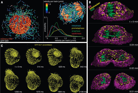
maybe the most important method in diagnosing illnesses and in biomedical research. The discipline is advancing in leaps and bounds. One of the most significant developers in the field is the Imaging Center at Janelia Campus, which is led by Chew.
Janelia Campus has built the Imaging Center and provided equipment that is not even available commercially. Researchers from all over the world can apply for a research period at the Center, during which Janelia covers all the expenses except food and travel and grants the laboratory to the exclusive use of the researcher.
One of the main ideas is to encourage scientists from other fields to use imaging in their research. For example, recently researchers have studied the bacteria found in oil, marine plants, the ink of an octopus, and death of coral with advanced microscopy.
– The idea is that now you can try out whatever you want. Think boldly. It is ok to fail, but if you do, do it impressively. Best science is achieved only through boldness, says Chew.
Global Network for Researchers
Chew has visited Turku several times and this time,
just before the opening seminar of the Bridging Nordic Imaging network, he teaches the newest imaging technology to the Master's degree students, doctoral candidates and researchers together with his colleague John Haddleston (in the photo).
– In recent years, bioimagign has become maybe the most important method in biomedical research in the whole world. Among other things, imaging includes new three-dimensional, super resolution microscopy techniques, non-invasive tissue and patient imaging methods, combinations of different methods used in diagnosing illnesses as well as computational analysis of results, describes Kankaanpää.
Chew continues that soon the development of the technology will make it possible for using the same tissue sample both in fluorescent and electron microscopy, as the sample will not have to be cut in its entirety before the imaging but only one slice at a time. In future, we can see inside living cells and individual molecules even better than at the moment.
– With imaging, we can observe a molecule in operation. How a cell supports life, how it creates diseases, how it functions. 
Chew and Kankaanpää stress the importance of co-operation. There is so much left to study in imaging that there is enough work for everyone. For example, in Finland the universities collaborate and decide together on their specialisation. The principle is open access – the researchers of other universities are welcome to use the equipment of the University of Turku and vice versa. However, the imaging community is small and the resources limited.
– All of us are involved in Finnish, Nordic European and global collaboration, says Kankaanpää.
Turku BioImaging Upholds Networks
Turku is quickly becoming one of the most central bioimaging centres in Europe. Turku leads some of the most important Finnish and international networks in the field.
Turku BioImaging was initiated by the University of Turku and Åbo Akademi University and received half a million Euro funding from the EU for making the new European imaging network, which is being established, open to researchers and for coordinating the sharing of expertise in a global imaging network and the principles of open access.
– The European model is a basis for Global BioImagine, says Chew.
The first link in the chain is the newly established Bridging Nordic Imaging network led by Turku BioImaging. Its first, strongly interdisciplinary seminar will bring the leading experts in imaging of the Nordic countries to Turku for 13–14 August. The topics of the seminar includes improving the infrastructure of Nordic imaging and presenting novel techniques that are under development, such as the photoacoustic imaging which is developed in Turku.
The recent upswing of Turku BioImaging is evident in several international top publications as well as in the Nobel Prize in Chemistry won by Stefan Hell in 2014 with his invention of super resolution microscopy, which he originally developed while working in Turku.
At the beginning of the week, Chew and his colleague, Applications Scientist John Heddleston, taught students and researchers of the Turku universities and they spend rest of the week attending the Bridging Nordic Imaging network's seminar, which was organised for the first time.
Text: Erja Hyytiäinen
Photos: Erja Hyytiäinen and Janelia
Translation: Mari Ratia