The Past, Present and Future of the Super Resolution STED Microscopy
– Without the University of Turku, my scientific career would never have continued, says Stefan Hell and ”raises” a glass of champagne via telephone to the crowd gathered in the break room of the Laboratory of Biophysics at the University of Turku.
Stefan Hell published the idea of the Stimulated Emission Depletion, i.e. STED, microscopy for the first time in 1994 when he was working in the University of Turku.
In addition to Erkki Soini, Kalervo Väänänen, Jyrki Heino and many other members of the University community, at the other end of the line Professor Pekkä Hänninen, Onkel Pekka as he is known to Hell’s children, also raises a glass. A week later, he relates to the audience at the Physics Day why this year’s Nobel Prize in Chemistry is also that of the University of Turku.
The auditorium at Quantum is filled to the last seat. The annual Physics Day usually shed more light on the winner of the Nobel Prize in Physics. This time, the amount of information is double: the work of the winners of the Nobel Prize in Chemistry, Eric Betzig, Stefan W. Hell and William E. Moerner, will be presented as well.
The introduction to the Nobel in Chemistry is given by Pekka Hänninen, Professor at the Institute of Biomedicine. The man who started working in collaboration with Hell in 1990 and, later together with Erkki Soini who at the time was a professor at the University of Turku, took part in developing the super resolution microscopy.
Ideas flew, some straight to the bin, others proceeded to development. One of the latter was an idea of the Stimulated Emission Depletion i.e. STED microscopy. Hell and Doctoral Candidate Jan Wichmann, who followed Hell to Turku from Germany, presented it in their publication in 1994.
– This is why we claim that this Nobel also belongs to Turku. The groundwork was done here, says Hänninen.
Rascals of Science
Pekka Hänninen strives to describe in popular terms to the audience what it is that Hell has done. However, first he has to start from Ernst Abbe who defined in 1873 in his theory of diffraction that the resolution limit of the traditional microscope is limited to 0.2 micrometers. This was long thought to be the cast-iron truth.
As a researcher in his late 20’s, Hänninen travelled to the European Molecular Biology Laboratory (EMBL) in Heidelberg to develop biological confocal microscopy. Confocal microscopes enable the study of three-dimensional structures by optically sectioning biological samples and were state-of-the-art at that time. Two years later, Stefan Hell joined the group to put his own ideas into practise.
However, Hell was considered a radical. Hänninen was warned against working with the man and he did not do so during the normal working hours. Yet, they happened to live in the same house, became friends and started working together after work in the evenings.
– Stefan was thought as ”the village idiot”, in a manner of speaking, and I guess that so was I, laughs Hänninen.
The Ties Did Not Break after Hell Returned to Germany
Hänninen returned to the University of Turku in 1992, but Hell’s work was coming to a sudden end. The controversial researcher did not find continuation for his funding even though he had already produced some results. It was fortunate for Hell – and to super resolution microscopy – that Hänninen convinced Professor Erkki Soini to give Hell a chance and the two men applied and were granted a funding to the joint project of Soini, Hänninen and Hell by the Academy of Finland.
–Stefan came to Turku at the beginning of 1993. The Academy of Finland was so curious that they exceptionally appointed a steering group to follow our work. None of the others who received funding had one, says Hänninen.
Ideas flowed. The researchers were convinced that the mindset where Abbe’s theory was “inviolable” was mistaken. They believed that the solution was to be found in quantum physics, in joint action of multiple photons in a sample. But how?
He talks in detail about the resolution of microscopes, stimulated emission, fluorescence, time delays, pulse trains and diffraction limit. He shows how STED microscopy uses two beams instead of one.
The first beam excites the molecules in the 0.2 micron focal spot and the other beam, shaped like an optical “doughnut”, depletes the fluorophores everywhere else but in the centre of the “doughnut”, a spot of few tens of nanometres, leaving these centre molecules to fluoresce. The image is then formed pixel by pixel on the computer when this tiny fluorescence spot of few tens of nanometres is moved over the biological sample through the area of interest.
–We had number of ideas. And something always worked producing new, sometimes unexpected results. When Stefan went back to Germany, optical nanoscopy was accepted only by a few, but he had made enough of a name with our “accidents” so that he could move forward, reminisces Hänninen.
Hell continued on his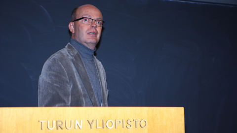
own path as a researcher and Hänninen (in the photo) on his own. Their last joint publication was published over a decade ago. The Laboratory of Biophysics continued with the application of the achieved results and examples of this are the TPX technology - virus and bacteria analysis of small children and Agsens technology for, quite surprisingly, water analysis in oil recovery.
However, the men are still in contact with each other. They became family friends and Hell has often been a visiting lecturer at the University of Turku, last time a year ago when the 20th anniversary of the invention of the super resolution microscopy was celebrated in Turku.
No Nobel without Applications
Hell continued his research in Germany. However, the microscope he created was not alone enough to win the prize. A Nobel also requires applications.
Hänninen reveals how STED 
(a STED microscope image on the right) has enabled the viewing of chemical phenomena in nerve impulses. It has been assessed that the work of Hell, Betzig and Moerner could enable researchers to find a cure for e.g. Alzheimer’s disease and osteoporosis. The most recent observation with the optical nanoscopy is how HIV infects a cell.
– We opened the door slightly, now the work is focusing in the applications, says Hänninen.
Text: Erja Hyytiäinen
Photos: Max Planck Institute for Biophysical Chemistry and Erja Hyytiäinen
Translation: Mari Ratia