The national Turku PET Centre is jointly run by the University of Turku, Åbo Akademi and the Hospital District of Southwest Finland and is situated right at the heart of the Turku University Hospital. The centre bustles with activity as five PET scanners constantly scan either patients or volunteers for research.
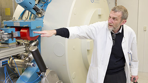
In the basement of the building, behind a two-metre thick concrete door is the centre of the whole operation: the particle accelerator.
– We received it from Russia as a payment for the loans of the USSR. The first particle accelerator of the PET Centre was acquired in 1974 by bilateral trading. In the trade, we got the particle accelerator, the USSR got eggs, reveals Professor of Radio Chemistry Olof Solin, who works at the PET Centre.
He explains in detail how the large magnet, a metre and a half in diameter, decays protons with 18 million volts of energy making their target nucleus unstable i.e. radioactive. The proton beam runs through different magnetic fields which direct it to the target materials.
– We mainly produce Carbon-11, Fluorine-18 and Copper-64, says Solin.
The nuclide map on the wall shows that there are dozens of nuclides and, in Turku, an unusually great amount of them can be produced, even on a worldwide scale. What this means is that the centre can diagnose multiple diseases and carry out exceptionally wide-ranging research.
Elevated Glucose Metabolism Reveals Cancer
Olof Solin has experienced the development and growth of the PET Centre. When Åbo Akademi acquired its first particle accelerator in 1974, Solin was writing his Master’s thesis and chose radionuclide production as his topic.
– We started researching short-lived radionuclides which are suitable for imaging, Solin reminisces back for nearly 40 years and then describes shortly in a nutshell what happens in positron emission tomography.
– The radionuclides produced with the particle accelerator decay due to positron emission. Positrons are the antiparticles of electrons, antimatter that cannot exist in our world. When a positron is generated, it immediately starts looking for an electron to interact with. Then the mass transforms into pure energy, into two gamma rays which are emitted in opposite directions, Solin describes the process.
The first particle accelerator was acquired for the research of nuclear physics but it was clear from the beginning that its capacity would be used for medical purposes as well. The researchers build the first imaging scanners themselves, the first actual PET scanner was acquired in 1988.
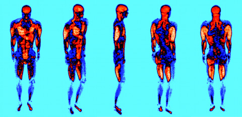
The PET image shows how the muscles function during cross-country skiing.
Solin tells how the PET scanner can recognise the decay process and define the trajectory of the quanta. With this information, the scanner can produce a precise image on, for example, where in the body the glucose metabolism is quick and where it is slower.
–For example, cancer tissue uses glucose as a fuel. When we have added Fluorine-18 to glucose and given the medicine to the patient, we can see with the scanner in which part of the body the glucose metabolism is elevated. The cancer tissue can be seen in the image as an active accumulation of glucose metabolism, says Solin.
Even Seconds Count in Different Stages of the Process
Only a few metres from the particle accelerator, which is protected by thick concrete, is the laboratory and, beyond that, a cleanroom which can only be entered by the personnel. An employee dressed in a blue protective coverall is at the moment combining a radionuclide created with the particle accelerator to a molecule.
–A radionuclide has to be combined with a molecule so that it forms a radiopharmaceutical. The molecule controls how the medicine is localised in the body. After we have combined the two, each batch of medicine is inspected is our laboratory. We analyse the solvent residue and the radiochemical and chemical purity. Only analysed and approved medicine will be given to the patients, says Solin.
At this stage of the process, each minute and, with some radiopharmaceuticals, even seconds are important.
– The half-life – i.e. the time it takes for half of the nuclides to have found an electron – of the used radionuclides ranges from two minutes to two hours. That’s why we have to produce the nuclides ourselves.
Half of the Capacity Used for Patients, the Other Half for Researc
On the upper floors there are five PET scanners. On a normal week day, each one of them is in a constant use. About half of the capacity is used to examining the health of individual patients and the other half is reserved for research.
– The original idea was to use 30 percent of the capacity for the treatment of patients, but that share has risen to about 50 percent. This is a national centre and we have responsibility to help patients from all around the country. It is the right thing to do and we are happy that we can provide clinical applications as well, says Solin.

The radioactivity of the radionuclides is one of the biggest challenges in the work. Therefore, remote-controlled synthesis methods have been developed for the laboratories; one of the newest methods uses robotics and was created by Doctor of Philosophy
Viki-Veikko Elomaa.
The applications are a result of devoted development work. The researchers of the PET Centre have managed to develop several drug ingredients that can be used to examine diseases that could not be detected with PET scanners before.
– Some of the research carried out with the scanners is our own basic research, whereas some of the research is for international pharmaceutical companies that have paid for it. All the leading pharmaceutical companies in the world have been in cooperation with us, says Solin revealing the respect that the centre has in the industry.
Part of the operations of the PET Centre is funded by the commercial financing received from the industrial cooperation. The basic funding comes from the universities and the hospital district and rest of the funding consists of academic research grants from the Academy of Finland and the EU.
PET Imaging Will Be Used to Reveal More Information about Diseases
At the PET Centre, researchers constantly develop new radiopharmaceuticals and methods of combining radionuclides with molecules. At the moment, for example, researchers are trying to find suitable radiopharmaceuticals for tracing the treatment of Alzheimer’s disease and diabetes.
– Another very interesting research is Jarmo Hietala’s attempt to find out with PET imaging what happens in the brain of a schizophrenic, says Solin.
The centre that started out with only a couple of employees now employs about 150 people.
– All the parties – the university, Åbo Akademi and the hospital district – have understood how valuable the PET Centre is and are ready to invest in it. We have also had very good directors, says Olof Solin.
The investment can be seen in the amount and quality of research.
– In the last few years, about 6 to 8 dissertations have been published each year and over a hundred peer-reviewed publications have been published, most of them in the top scientific journals of the world, says Solin.
Text: Erja Hyytiäinen
Photos: Antti Tarponen and PET image from publication: Bojsen-Møller J, Losnegard T, Kemppainen J, Viljanen T, Kalliokoski KK, Hallén J. Muscle use during cross country skiing evaluated by positron emission tomography. Journal of Applied Physiology, 109(6):1895-1903, 2010.
Translation: Mari Ratia