Valuable Imaging Equipment for Shared Use at Turku BioImaging
Turku BioImaging offers its researchers a cost-effective possibility to versatile biomedical imaging. The crown jewel of the centre is a brand new super-resolution STED microscope which is the only one in Finland. All operations aim to bring the valuable equipment to as efficient as possible public use.
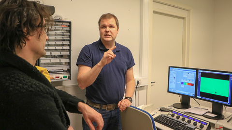
Postdoctoral Researcher Ilkka Paatero (on the right) presented the multiphoton microscopy. According to biochemistry researcher Anssi Malinen (on the left), who was visiting the facilities, the diverse imaging equipment of the Turku BioImaging enables versatile cell level research programmes.
– When procuring new equipment, research groups consider together which device is needed. One applies, and others support the application. If funding is received, the device will be placed in the BioImaging facilities, and for an operating charge other research groups can also use it, said Graduate School Coordinator Markku Saari while presenting the equipment in the Open Day at the Turku BioImaging.
This open access principle beats the old way in which successful professors received funding for a piece of imaging equipment that was then locked up so that no one else had access to it. According to Chair of Turku BioImaging John Eriksson, this new way of thinking has made it possible to collect all the different imaging technologies needed on campus in the shared facilities.
– We have a web-based booking system, clear instructions and a price list. The price paid by a researcher has been strongly subsidised, since external funding has been received for the procurement costs. In the long run, the open access principle is extremely cost-effective, says Eriksson.
Those conducting academic research may pay 20 euros per hour for the use of a device, whereas with a company, the price is negotiated on a case-by-case basis but will be multifold as a rule of thumb.
Eriksson explains how a user of a device must complete a device-specific training to get a permit. If the researcher does not have a permit, the centre provides an expert to help for an additional compensation. The expert will go through the samples with the researcher so that they get exactly the pictures they want.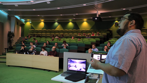
The head of the Finnish Euro-Bioimaging Node Camilo Guzman introduced the services of Turku BioImaging to visitors. The centre is a part of the European Euro-BioImaging (EuBI) imaging network that offers researchers a possibility to use the modern imaging technologies they need in different parts of Europe.
– First and foremost, we want as many as possible to learn how to use the equipment, says the head of the Finnish Euro-Bioimaging Node Camilo Guzman.
Part of European Network

Graduate School Coordinator Markku Saari presented different types of microscopes during the Open Day. The equipment is available to different researchers, and according to Saari, researchers can be found working with the equipment to late in the evening and during the weekend.
The task of the nodes in the EuBI organisation is to guide and help researchers in finding the optimal solutions for research questions related to biomedical imaging and make top technologies available to them.
Until recently, the equipment used for imaging have required great investments from the research groups and universities. Whereas now, all researchers can use the needed technologies through the network, even if the necessary equipment is not available at their home university.
– Many users have an idea of how they could solve a research problem but cannot advance their research without the required equipment. Other users, then again, struggle with scientific problems and are unaware of the available possibilities that could be used for solving the problems, says Guzman.
Bioimaging includes a broad spectrum of biomedical research techniques, such as microscopy techniques, and magnetic resonance and PET imaging. These techniques enable the application of laboratory-based basic research, for example, to developing new medicines or patient diagnostics, and help the development of medical breakthroughs.
>> Turku BioImaging
Text: Erja Hyytiäinen
Translation: Saara Yli-Kauhaluoma