A new doctoral dissertation shows that gambling disorder is linked to brain networks involved in self-control and brain reward functions. By combining several brain imaging methods, the research provides new biological insight into the disorder and may point to promising directions for treatment development.
Gambling disorder is a condition in which gambling becomes difficult to control and causes harm to well-being, relationships, and daily functioning. Gambling disorder affects around 1–2% of adults worldwide and, due to its similarities with substance addictions, it is the first behavioral addiction officially recognized in diagnostic manuals.
In his dissertation research, Doctoral Researcher Albert Bellmunt Gil from the University of Turku in Finland aimed to better understand the brain abnormalities in gambling disorder, which can provide insight into why gambling behavior persists in affected individuals despite even serious negative consequences.

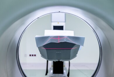
The research focused on investigating the frontal lobe and striatum, areas of the brain that regulate self-control, decision-making, and brain reward functions. The research was conducted in two independent datasets including people with gambling disorder and healthy control participants, using several brain imaging techniques that measure brain structure, brain activity, and chemical signaling.
Brain imaging reveals altered control and reward circuits
The results showed that gambling disorder is linked to disrupted connections between the frontal cortex and subcortical brain regions, i.e., fronto-striatal circuits.
“In particular, connections between the dorsolateral frontal cortex and nucleus accumbens – the key brain reward nucleus – were weaker than normal, which may make it harder to stop gambling once urges arise,” says Bellmunt Gil.
Compared to healthy volunteers, people with gambling disorder also showed a stronger brain response to gambling-related cues in the dorsal striatum, paralleling earlier findings in individuals with substance use disorders to cues associated with drug use.
Abnormal fronto-striatal connectivity was associated with brain serotonin and cue-reactivity to brain opioid function.
“People with gambling disorder also had brain structural abnormalities within the fronto-striatal circuit, which may represent an underlying vulnerability to developing gambling disorder or they are caused by long-term excessive gambling,” Bellmunt Gil explains.
Toward more targeted treatments
The findings provide biological evidence that may help guide more targeted treatment approaches in the future.
“For example, the findings highlight brain regions that are already being targeted with therapy in other conditions, such as non-invasive brain stimulation, which uses magnetic pulses to influence brain activity. The fronto-striatal circuit identified in this study provides a testable target for non-invasive brain stimulation for gambling disorder,” explains Bellmunt Gil.
In addition, the findings related to serotonin and opioid function suggest that targeting these neurotransmitters with medications might be beneficial. However, randomised controlled studies are needed to test these hypotheses.
“Our results show that gambling disorder is associated with measurable changes in brain areas that regulate control, reward, and habits,” says the researcher. “Understanding these brain mechanisms can help reduce stigma and support the development of more effective treatments.”
The findings of this dissertation study reinforce the idea that gambling disorder is not a matter of willpower, but a condition linked to changes in brain function and structure.
Improved understanding of these brain mechanisms may support better prevention strategies, more effective treatments, and a more compassionate view of people affected by gambling disorder.
For more information, please contact: abegil@utu.fi