Turku can, for a good reason, be called the imaging capital of Finland and even the entire Europe. The city is home to both the headquarters of the Euro-BioImaging research infrastructure and the national Turku PET Centre, one of the continent’s most important medical imaging centres.
Imaging refers not only to medical imaging techniques, such as PET and MRI, but also to advanced microscopy techniques, which, with the aid of modern optics, lasers, and software, make it possible to see in real time how a virus penetrates a cell or analyse the movement of cancer cells, for instance.
Turku has a long history in the development of imaging techniques and has acted as an example, even on the international arena, in making imaging available to everyone who needs it.
“Euro-BioImaging is a pan-European imaging infrastructure that consists of nodes, or service centres, in different countries, where researchers can come to conduct their research. If a researcher cannot find necessary technology in their own university or company, they can come to any Euro-BioImaging service centre,” says Tiina Saanijoki, Medical Imaging Research Manager at Turku BioImaging.
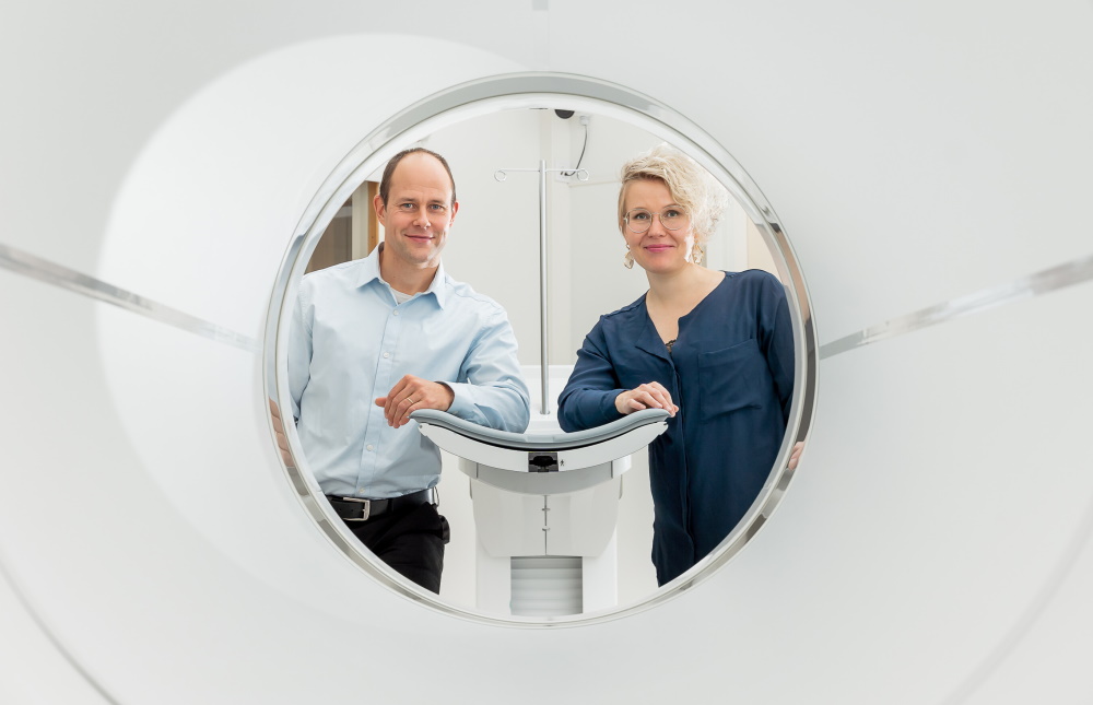
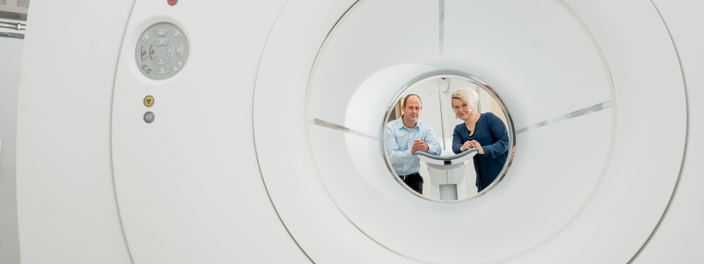
In their research, Riku Klén and Tiina Saanijoki are focusing especially on PET imaging.
Saanijoki works as the coordinator of Euro-BioImaging’s Finnish Biomedical Imaging Node. In her own research, she has used PET imaging to study pleasure that physical activity generates in the brain.
In Finland, six universities and three university hospitals are part of Euro-BioImaging, making it one of the most popular imaging service clusters in Europe. Imaging technology is expensive and requires an extremely high level of expertise. Euro-BioImaging’s goal is to make most important technologies available under single window service.
“Broad infrastructure makes it possible to define the research question more freely as the tools for finding answers are available to everyone. This, in turn, yields better and more useful research results,” Saanijoki points out.
Bioimaging makes pharmaceutical development possible
Euro-BioImaging and the national Turku PET Centre offer services to both researchers and private sectors such as the pharmaceutical industry. Openly available imaging services also improve smaller companies’ possibilities to conduct research as they do not need to have expensive equipment and special expertise themselves. For instance, Turku PET Centre has collaborated for a long time with both international pharma giants and local start-ups.
Bioimaging is one of the most important tools in biomedicine, on which nearly all modern pharmaceutical development and other research in the field rely at one development stage or another. Imaging enables research on pathogenesis (disease development mechanisms) and disease background factors. It can also be used for identifying various medication targets, such as which cells are targeted with pharmaceuticals, how different viruses affect cells or which protein yields the desired effects on cells.
"Imaging can reveal if the candidate molecule bonds with the desired target and how strong its therapeutic effect is."
– Tiina Saanijoki
“When the target is identified, there are drug candidate molecules, which are studied for the desired effect. Imaging can reveal if the candidate molecule bonds with the desired target and how strong its therapeutic effect is. We can also find out how a molecule distributes within the body and whether it finds its way to the intended location or remains in the circulation, for instance. One could say that imaging makes invisible visible,” Saanijoki explains.
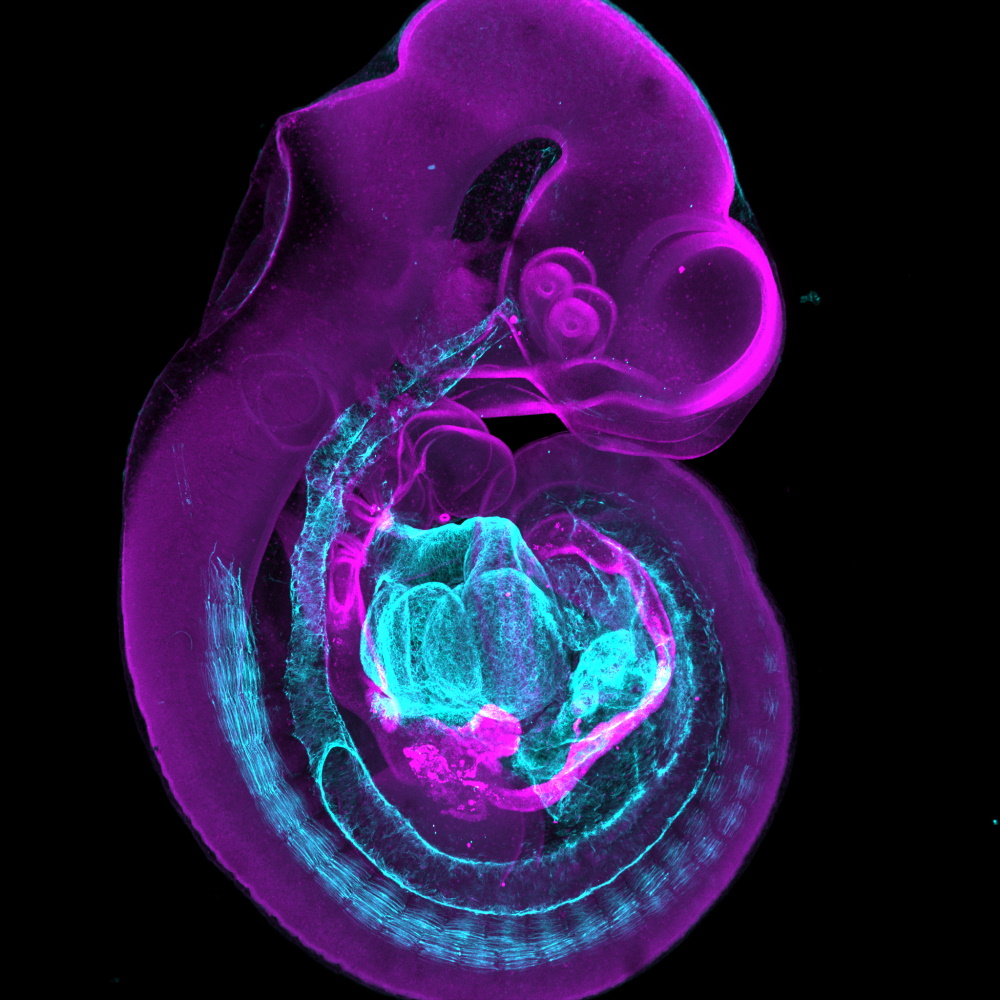
Animal experiments provide important information how diseases develop and are a key element in early-stage drug development. The image shows a mouse embryo scanned with a stereomicroscope. Image: Ciarán Butler-Hallissey (Turku Bioscience Centre).
According to Saanijoki, world-class bioscientific, medical and biotechnological research is conducted in Turku partly because we have invested in state-of-the-art research equipment and expertise, but also because excellent cohort materials, a multidisciplinary university, and important international contacts provide brilliant preconditions for both high-quality basic research and treatment-related research.
“In addition, Turku PET Centre has a unique offering of radiopharmaceuticals, which sets us apart from other PET research institutes also globally. Turku also has top expertise in advanced light microscopy, image analysis, mathematics, and artificial intelligence, for instance, which makes a wide range of research questions possible.”
A total-body PET scanner opens up new research and treatment opportunities
A recent example of Turku’s exceptional research infrastructures is a total-body PET scanner, which was taken into use at the national Turku PET Centre last spring.
“The introduction of new equipment always requires the development of new methods, too. Today, it requires expertise in mathematics and software development,” says Riku Klén, Assistant Professor at Turku PET Centre.
"The introduction of new equipment always requires the development of new methods, too."
– Riku Klén
Klén’s original field of study was mathematics, and now his research team is focused on making the best use of the total-body PET scanner. The team develops imaging methods for the new PET scanner, image modelling, and different types of software related to the use of the equipment. Imaging generates so much data that data structuring and analysis requires artificial intelligence, in other words, algorithmic data management and machine learning methods.
“The total-body PET scanner holds enormous potential for research and diagnostics. The traditional PET scanners have only been able to scan 20–25cm slices of the subject at a time. We are now on the brink of a transformation: the total-body PET scanner makes it possible to carry out the simultaneous imaging of the brain and the heart, for instance, which enables new kinds of research designs and better treatment in health care services. If a patient comes to the hospital with chest pain, it could be a heart attack but the problem might also be in the brain or another part of the body. With the new PET scanner, we see a full-body image at one go. For patients, the new device means a lower amount of radiation and a shorter scanning time.”
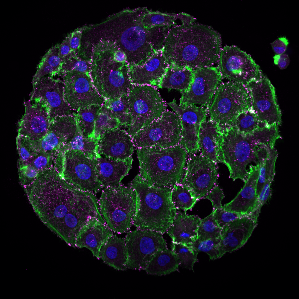
Spreading of breast cancer cells can be imaged using a spinning disk confocal microscope. Image: Guillaume Jacquemet (Åbo Akademi)
According to Klén, his team also co-operates closely with the medical researchers of Turku University Hospital. The team’s goal is to create clinical tools to ensure better diagnostics and treatment.
“For instance, we have developed a new kind of accelerometer-based measuring device for PET imaging, capable of monitoring the movements of the heart and the lungs and compensating for the movement-induced imprecision in images.”
Text: Heikki Kettunen
Images: Hanna Oksanen, Ciarán Butler-Hallissey & Guillaume Jacquemet
The article has been published in the Aurora magazine’s issue 2/2022.