The University of Turku, the University of Münster and the University Medical Center Utrecht are developing new solutions for online teaching based on visual material. During the EU-sponsored project, which will run until 2023, new digital tools coupled with innovative pedagogical scenarios will be tested in the teaching of pathology. At the University of Turku, there are researchers from the Faculty of Education and the Faculty of Medicine involved in the project.
The cLovid project is developing digital tools to facilitate online teaching and learning in disciplines that are strongly based on the use of visual material in teaching. For instance, students are able to make place markers or annotations on the material presented, and the information can be shared with other students and the teacher.
– Zoom is used in online teaching widely, but up til now the students have had limited possibilities to interact with the visual material and share their observations by referring to exact coordinates in the material. We will integrate these functions. In the future, students can mark their observations on the screen and after marking them, they can discuss them together. In cLovid, we are also testing the usability of the programs in medical education, says University Research Fellow, Dr, Laura Helle from the Department of Teacher Education at the University of Turku.
In the consortium, Turku's role is to plan and carry out naturalistic teaching experiments; in addition, researchers from the University of Turku will evaluate the development process and its results. In addition to Dr. Helle, project researcher Mikko Kainulainen from the Department of Teacher Education and Docent Pauliina Kronqvist from the Department of Biomedicine are working on the project.
The development work is funded by Erasmus + strategic partnership funding from the EU. The project was launched in 2021 and will run until 2023. According to Helle the impetus for the project was an extra call for funding motivated by the COVID-19 pandemic. However, the basis is long-standing development work. Koen Vincken from University Medical Center Utrecht and Bas de Leng from University of Münster are among the best in their field. Vincken specializes in medical imaging applications and de Lengin nnovative medical learning materials and environments. de Leng is the coordinator of the cLovid project.
The development work is funded by the EU
When the EU decided to allocate special funding to cope with exceptional circumstances like pandemics in the future, and at the same time the pandemic made it difficult to teach microscopy of pathology, the pieces fell into place.
– The corona blockage resulted in a digital leap in microscopy teaching: a large amount of teaching was recorded and made available to the students. What was missing was interactivity and feedback for an individual student, says clinical teacher, docent Pauliina Kronqvist from the Department of Biomedicine, University of Turku.
The cLovid project focuses primarily on basic education in pathology as part of undergraduate education in medicine, but is also interested in exploring the wider applicability of the tools developed.
– Could the applications of digital microscopy be useful in the teaching of biology, for example, Helle ponders. - The advantage is that the whole class can “examine” one sample either at the same time or even in advance. The findings and conclusions can then be discussed together.
At the heart of the development work are two pieces of software
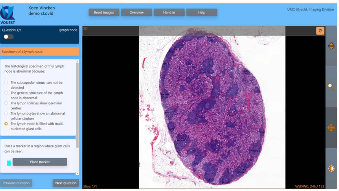
At the heart of the cLovid project development there are two pieces of software: VQuest and PRISMA. In addition, an open source “virtual microscope” or microscopic specimen viewer is being developed further.
Originally, conceived over a decade ago, VQuest was originally developed to meet the needs of radiology specialist training. It is utilized, e.g., in annual progress tests in Utrecht assessing the skills of Dutch radiology residents.
With VQuest, students can practice their image interpretation skills, and teachers can even get real-time feedback on student performance. This allows the teacher to pass on feedback to students, even in the case of large group teaching.
PRISMA is newer piece of software. PRISMA is a platform designed to present the results of interpretations and analyses visual materials in formats that promote interactive and reflective learning. With PRISMA, teachers can produce summaries of the results of tasks performed on VQuest, for example, and open a discussion about critical issues or misunderstandings.

– The aim of the project is to pilot the use of the latest software and educational solutions in online teaching of pathology. The solutions can be utilized in both distance and face-to-face teaching, by utilizing, for example, students' own laptops, Helle says.
– In recent years, microscopy teaching, which is part of basic medical education, has become almost completely digital. Teaching is accomplished by digitizing cell and tissue samples “piece by piece” into large image files that are viewed with a virtual microscope, says Kronqvist.
From the University of Turku, there are researchers from the Faculties of Education and Medicine involved. The project is multidisciplinary and its core group has expertise in learning research, pathology, medical informatics and teaching technology.
– Although the project involves several people with solid multidisciplinary competence, none of us would be able to do this development work alone. Interdisciplinarity also poses everyday challenges: especially in the early stages of the project, the most challenging thing was to understand what each researcher was talking about, Helle says.
Pathology and anatomy teachers test tools in teaching
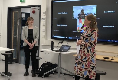
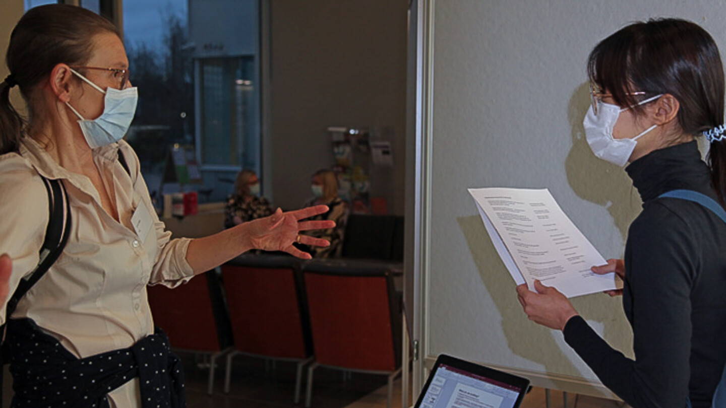
At the end of May, the cLovid project organized a two-day seminar in Amsterdam, bringing together a group of pathology and anatomy teachers, medical students, learning researchers and medical software experts. The seminar had several objectives.
– We wanted to introduce clinical teachers to the software and teaching methods that are still being developed. Through this, we sought both feedback on the development work and ideas for piloting with different teachers, says project coordinator Bas De Leng from the University of Münster.
Based on the work done in Amsterdam, several pilots are now planned. These are carried out together with clinical teachers from the universities of Turku, Maastricht, Oulu and Eastern Finland.

At the Amsterdam seminar, small groups developed blended learning scenarios for pathology teaching . Pictured (from left) are Sönke Scherzer, Friedrich Pawelka, Bas de Leng, Katarina Korpinen, Pauliina Kronqvist, Jack Cleutjens, Hannie Gijlers and Koen Vincken. Photo: Mikko Kainulainen.
The creation of these resources has been (partially) funded by the ERASMUS+ grant program of the European Union under grant no. 2020-1-DE01-KA226-HE-005813.